Отдел лучевой диагностики


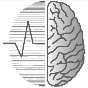
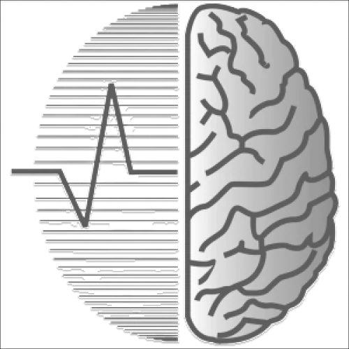
Отделение лучевой диагностики Научного центра неврологии стало пионером применения компьютерной томографии в нашей стране. Разработана методология пошаговых КТ-морфологических сопоставлений. Сотрудникам отделения совместно с инженерами и физиками в 1984 году была присуждена Государственная премия за создание отечественной модели рентгеновского компьютерного томографа. В отделении разработаны оригинальные технологии совмещения изображений, получаемых на компьютерных томографах различных типов (КТ, МРТ, ОФЭКТ). Разработан комплекс нейровизуализационных биомаркеров нейродегенеративных, демиелинизирующих и цереброваскулярных заболеваний, включающий оценку ряда морфометрических, трактографических и функциональных параметров и направленный на объективный мониторинг течения болезни, в том числе на фоне проводимой терапии. Проведено функциональное картирование речевых и двигательных зон головного мозга с целью определения реабилитационного потенциала и выработки персонифицированных протоколов восстановительного лечения пациентов с инсультом.
ОСНОВНЫЕ НАПРАВЛЕНИЯ ДЕЯТЕЛЬНОСТИ
Основной профиль отделения – нейровизуализация, работа ведется в двух направлениях: клиническом и научно-исследовательском. Основные задачи отделения: исследование строения мозга в норме и при патологии, физиологии и патофизиологии заболеваний центральной нервной системы (ЦНС) диагностика сложных случаев поражения ЦНС, прогнозирование течения заболеваний, определение реабилитационного потенциала больных, контроль эффективности проводимой терапии и др. разработка алгоритмов исследования пациентов с заболеваниями ЦНС. Клиническая нейровизуализация: применение МРТ и МСКТ в диагностике сосудистых (ишемических инсультов, внутримозговых кровоизлияний), инфекционных, рассеянного склероза, других демиелинизирующих, нейродегенеративных заболеваний, опухолей ЦНС и других заболеваний центральной нервной системы. обследование пациентов с заболеваниями позвоночника, межпозвонковых дисков и связочного аппарата (включая функциональные пробы, в том числе динамическую МРТ шейного отдела позвоночника). МРТ-исследование поражений черепных нервов, нервных сплетений и нервов при периферических нейропатиях. МРТ-диагностика нейромышечных заболеваний. функциональное картирование коры головного мозга перед нейрохирургическими вмешательствами (моторные, речевые парадигмы фМРТ и проч.). рентгеноскопия акта глотания при дисфагиях различного генеза Научно-исследовательская работа: КТ- и МРТ-перфузия головного мозга при различных патологических состояниях ЦНС, включая методику 4D-adaptive одновременного получения ангиографии, веносинусографии и перфузии мозга. Оценка кровотока по магистральным артериям головы и шеи, оценка структуры стенки сосудов при их поражении (васкулиты, атеросклероз, диссекция). Сочетание различных МРТ-методик в оценке патогенеза и прогрессирования болезни малых сосудов. Мультимодальное МР-исследование рассеянного склероза и других демиелинизирующих заболеваний (диагностика, оценка прогрессирования процесса, прогнозирование функционального исхода). Нейровизуализационные аспекты гетерогенности инфарктов в полушариях большого мозга. Функциональная МРТ как в норме (картирование), так и в изучении пластичности головного мозга при различной патологии ЦНС, в качестве маркера восстановления нарушенных функций ЦНС. Кроме того, методика активно используется для выявления функционально значимых зон при планировании хирургических вмешательств на головном мозге. Диффузионные методы исследования: диффузионно-взвешенная МРТ и диффузионно-тензорная МРТ. Последняя применяется для выявления изменений проводящих путей головного мозга при сосудистых, дегенеративных и демиелинизирующих заболеваниях. Данные методики позволяют оценивать степень вовлечения белого вещества в патологический процесс и прогнозировать течение заболевания и потенциал восстановления пациентов. МРТ-морфометрия в изучении ультраструктурных изменений головного мозга при различных заболеваниях ЦНС. МРТ-спектроскопия в дифференциальной диагностике поражений нервной системы. Мультимодальное картирование головного мозга (сочетание методик структурной МРТ, фМРТ, ДТ-МРТ, ЭЭГ, ТМС). Новый подход к оценке физиологических параметров деятельности головного мозга при совмещении изображений, получаемых с помощью различных методов нейровизуализации.
УЧЕБНО-МЕТОДИЧЕСКАЯ РАБОТА
На базе отделения лучевой диагностики реализуются программы высшего образования по подготовке в ординатуре по специальности 31.08.09 Рентгенология (срок обучения - 2 года), в аспирантуре по направлению 31.06.01. Клиническая медицина, направленность «Лучевая диагностика, лучевая терапия» (очная форма, срок обучения - 3 года). Также на базе отделения лучевой диагностики (группа КТ и МРТ) проводятся курсы повышения квалификации врачей-рентгенологов по теме: "КТ и МРТ в диагностике заболеваний ЦНС". По всем вопросам обращаться в Ученую часть ФГБНУ НЦН тел.: 8(495) 490-21-04
ОБОРУДОВАНИЕ В СОСТАВЕ ОТДЕЛЕНИЯ
Магнитно-резонансные томографы закрытого типа: МР-томограф нового поколения Magnetom Prisma SIEMENS (Германия) – величина магнитной индукции 3 Тесла Magnetom Verio, Siemens (Германия) – величина магнитной индукции 3 Tесла, диаметр апертуры – 70 см, вес пациента – до 130 кг; Magnetom Avanto, Siemens (Германия) – величина магнитной индукции 1.5 Tесла, диаметр апертуры – 60 см, вес пациента – до 130 кг; Магнитно-резонансные томографы открытого типа: Magnetom C!, Siemens (Германия) – величина магнитной индукции 0.4 Tесла, вес пациента – до 130 кг; Panorama, Philips (Нидерланды) – величина магнитной индукции 1.0 Tесла, вес пациента - до 130 кг. Мультиспиральные компьютерные томографы: Somatom Definiton AS+, Siemens, (Германия) - 128-срезовый Цифровой рентгенодиагностический аппарат Italrhoentgen Clinodigit (Италия); Денситометр Stratos dR DMS group (Франция).
- Магниторезонансная томография
- Рентгеновская компьютерная томография
Топ-100
Области наук
Журналы


Цитирующие журналы


Цитируемые журналы


Издатели
|
5
10
15
20
25
|
|
|
Media Sphere Publishing House
23 публикации, 19.66%
|
|
|
Scientific Center of Neurology
14 публикаций, 11.97%
|
|
|
Springer Nature
10 публикаций, 8.55%
|
|
|
Pleiades Publishing
9 публикаций, 7.69%
|
|
|
MDPI
9 публикаций, 7.69%
|
|
|
ANO NCR
8 публикаций, 6.84%
|
|
|
Pirogov Russian National Research Medical University
6 публикаций, 5.13%
|
|
|
Elsevier
4 публикации, 3.42%
|
|
|
Frontiers Media S.A.
4 публикации, 3.42%
|
|
|
Eco-Vector LLC
4 публикации, 3.42%
|
|
|
Central Research Institute of Radiation Diagnostics
4 публикации, 3.42%
|
|
|
Wiley
3 публикации, 2.56%
|
|
|
Luchevaya Diagnostika
2 публикации, 1.71%
|
|
|
IMA Press, LLC
2 публикации, 1.71%
|
|
|
Moscow Regional Research and Clinical Institute (MONIKI)
2 публикации, 1.71%
|
|
|
Vidar, Ltd
2 публикации, 1.71%
|
|
|
Baltic Medical Education Center
1 публикация, 0.85%
|
|
|
Silicea - Poligraf, LLC
1 публикация, 0.85%
|
|
|
American Psychological Association (APA)
1 публикация, 0.85%
|
|
|
Publishing House ABV Press
1 публикация, 0.85%
|
|
|
The Russian Academy of Sciences
1 публикация, 0.85%
|
|
|
Endocrinology Research Centre
1 публикация, 0.85%
|
|
|
Medical Informational Agency Publishers
1 публикация, 0.85%
|
|
|
Paediatrician Publishers LLC
1 публикация, 0.85%
|
|
|
Geotar-Media Publishing Group
1 публикация, 0.85%
|
|
|
International Society for Neurofeedback Research (ISNR)
1 публикация, 0.85%
|
|
|
Federal Research and Clinical Center for Resuscitation and Rehabilitation
1 публикация, 0.85%
|
|
|
5
10
15
20
25
|
Организации из публикаций
|
10
20
30
40
50
60
70
80
90
|
|
|
Российский центр неврологии и нейронаук
81 публикация, 69.23%
|
|
|
Организация не определена
|
Организация не определена, 33, 28.21%
Организация не определена
33 публикации, 28.21%
|
|
Московский государственный университет имени М.В. Ломоносова
9 публикаций, 7.69%
|
|
|
Национальный исследовательский университет «Высшая школа экономики»
4 публикации, 3.42%
|
|
|
Башкирский государственный медицинский университет
4 публикации, 3.42%
|
|
|
Российский национальный исследовательский медицинский университет имени Н. И. Пирогова
4 публикации, 3.42%
|
|
|
Научно-технологический университет «Сириус»
3 публикации, 2.56%
|
|
|
Научный Центр Психического Здоровья
3 публикации, 2.56%
|
|
|
Медицинская школа имени Чана при Массачусетском университете
3 публикации, 2.56%
|
|
|
Институт общей генетики им. Н.И. Вавилова РАН
2 публикации, 1.71%
|
|
|
Университет Осло
2 публикации, 1.71%
|
|
|
Национальный исследовательский центр "Курчатовский институт"
1 публикация, 0.85%
|
|
|
Институт медико-биологических проблем РАН
1 публикация, 0.85%
|
|
|
Национальный медицинский исследовательский центр гематологии
1 публикация, 0.85%
|
|
|
НМИЦ нейрохирургии им. академика Н.Н. Бурденко Минздрава России
1 публикация, 0.85%
|
|
|
Университетская клиника Осло
1 публикация, 0.85%
|
|
|
Университет Мюнстера
1 публикация, 0.85%
|
|
|
10
20
30
40
50
60
70
80
90
|


Страны из публикаций
|
10
20
30
40
50
60
70
80
90
|
|
|
Россия
|
Россия, 87, 74.36%
Россия
87 публикаций, 74.36%
|
|
Страна не определена
|
Страна не определена, 33, 28.21%
Страна не определена
33 публикации, 28.21%
|
|
США
|
США, 5, 4.27%
США
5 публикаций, 4.27%
|
|
Норвегия
|
Норвегия, 4, 3.42%
Норвегия
4 публикации, 3.42%
|
|
Германия
|
Германия, 1, 0.85%
Германия
1 публикация, 0.85%
|
|
Саудовская Аравия
|
Саудовская Аравия, 1, 0.85%
Саудовская Аравия
1 публикация, 0.85%
|
|
10
20
30
40
50
60
70
80
90
|
Цитирующие организации


Цитирующие страны
- Мы не учитываем публикации, у которых нет DOI.
- Статистика пересчитывается раз в сутки.
