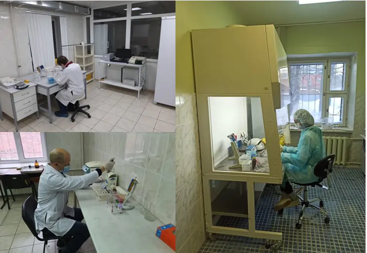
Центральная научно-исследовательская лаборатория


Центральная научно-исследовательская лаборатория (ЦНИЛ) Омской государственной медицинской академии создана по приказу МЗ РСФСР в 1964 году как научное подразделение в целях создания единого межкафедрального научно-методического и экспериментально-клинического центра ВУЗа для проведения комплексных работ по наиболее актуальным медико-биологическим проблемам. Вся деятельность лаборатории связана с научной и учебной работой академии. С учетом приоритетных направлений научно-исследовательской работы, сложившихся в научных коллективах академии, в 2001 г. проведена реорганизация ЦНИЛ с выделением отделов: - отдела экспериментальной медицины; - отдела молекулярно-биологических методов, который состоит из следующих самостоятельных подразделений: - иммунологическая лаборатория; - биохимическая лаборатория; - радиологическая лаборатория - лаборатория клинической микробиологии; - ПЦР-группа.
- Иммуноферментный анализ (ИФА)
- ПЦР
- ПЦР-РВ
- Различные методы молекулярной биологии

Топ-100
Области наук
|
1
2
3
4
|
|
|
General Medicine
|
General Medicine, 4, 22.22%
General Medicine
4 публикации, 22.22%
|
|
Obstetrics and Gynecology
|
Obstetrics and Gynecology, 3, 16.67%
Obstetrics and Gynecology
3 публикации, 16.67%
|
|
Materials Chemistry
|
Materials Chemistry, 2, 11.11%
Materials Chemistry
2 публикации, 11.11%
|
|
Gastroenterology
|
Gastroenterology, 2, 11.11%
Gastroenterology
2 публикации, 11.11%
|
|
Metals and Alloys
|
Metals and Alloys, 1, 5.56%
Metals and Alloys
1 публикация, 5.56%
|
|
Surfaces, Coatings and Films
|
Surfaces, Coatings and Films, 1, 5.56%
Surfaces, Coatings and Films
1 публикация, 5.56%
|
|
Organic Chemistry
|
Organic Chemistry, 1, 5.56%
Organic Chemistry
1 публикация, 5.56%
|
|
Water Science and Technology
|
Water Science and Technology, 1, 5.56%
Water Science and Technology
1 публикация, 5.56%
|
|
Hepatology
|
Hepatology, 1, 5.56%
Hepatology
1 публикация, 5.56%
|
|
Forestry
|
Forestry, 1, 5.56%
Forestry
1 публикация, 5.56%
|
|
Geography, Planning and Development
|
Geography, Planning and Development, 1, 5.56%
Geography, Planning and Development
1 публикация, 5.56%
|
|
General Earth and Planetary Sciences
|
General Earth and Planetary Sciences, 1, 5.56%
General Earth and Planetary Sciences
1 публикация, 5.56%
|
|
Economics and Econometrics
|
Economics and Econometrics, 1, 5.56%
Economics and Econometrics
1 публикация, 5.56%
|
|
Pediatrics, Perinatology and Child Health
|
Pediatrics, Perinatology and Child Health, 1, 5.56%
Pediatrics, Perinatology and Child Health
1 публикация, 5.56%
|
|
Media Technology
|
Media Technology, 1, 5.56%
Media Technology
1 публикация, 5.56%
|
|
1
2
3
4
|
Журналы
|
1
2
3
|
|
|
Russian Bulletin of Obstetrician-Gynecologist / Rossiyskii Vestnik Akushera-Ginekologa
3 публикации, 16.67%
|
|
|
Medical alphabet
2 публикации, 11.11%
|
|
|
Voprosy Prakticheskoi Pediatrii
1 публикация, 5.56%
|
|
|
Protection of Metals and Physical Chemistry of Surfaces
1 публикация, 5.56%
|
|
|
Pediatriya - Zhurnal im G.N. Speranskogo
1 публикация, 5.56%
|
|
|
Tuberculosis and Lung Diseases
1 публикация, 5.56%
|
|
|
Medical News of North Caucasus
1 публикация, 5.56%
|
|
|
Perm Medical Journal
1 публикация, 5.56%
|
|
|
Clinical Medicine (Russian Journal)
1 публикация, 5.56%
|
|
|
Russian Journal of Evidence-Based Gastroenterology / Dokazatelnaya Gastroenterologiya
1 публикация, 5.56%
|
|
|
Experimental and Clinical Gastroenterology
1 публикация, 5.56%
|
|
|
Pharmateca
1 публикация, 5.56%
|
|
|
Сибирский вестник психиатрии и наркологии
1 публикация, 5.56%
|
|
|
Kazan medical journal
1 публикация, 5.56%
|
|
|
Transbaikalian Medical Bulletin
1 публикация, 5.56%
|
|
|
1
2
3
|


Цитирующие журналы
|
1
2
3
|
|
|
Bulletin of Siberian Medicine
3 цитирования, 15.79%
|
|
|
Protection of Metals and Physical Chemistry of Surfaces
3 цитирования, 15.79%
|
|
|
Medical Immunology (Russia)
3 цитирования, 15.79%
|
|
|
Medical Journal of the Russian Federation
2 цитирования, 10.53%
|
|
|
Gigiena i sanitariia
1 цитирование, 5.26%
|
|
|
Tuberculosis and Lung Diseases
1 цитирование, 5.26%
|
|
|
AIP Conference Proceedings
1 цитирование, 5.26%
|
|
|
Meditsinskiy sovet = Medical Council
1 цитирование, 5.26%
|
|
|
Acta Biomedica Scientifica (East Siberian Biomedical Journal)
1 цитирование, 5.26%
|
|
|
Clinical Medicine (Russian Journal)
1 цитирование, 5.26%
|
|
|
Annals of critical care
1 цитирование, 5.26%
|
|
|
The actual problems in dentistry
1 цитирование, 5.26%
|
|
|
1
2
3
|


Цитируемые журналы


Издатели
|
1
2
3
4
|
|
|
Media Sphere Publishing House
4 публикации, 22.22%
|
|
|
Eco-Vector LLC
2 публикации, 11.11%
|
|
|
Alfmed LLC
2 публикации, 11.11%
|
|
|
Pleiades Publishing
1 публикация, 5.56%
|
|
|
Dynasty Publishing House
1 публикация, 5.56%
|
|
|
Stavropol State Medical University
1 публикация, 5.56%
|
|
|
Bionika Media Innovations
1 публикация, 5.56%
|
|
|
Medical Informational Agency Publishers
1 публикация, 5.56%
|
|
|
Mental Health Research Institute
1 публикация, 5.56%
|
|
|
Chita State Medical Academy
1 публикация, 5.56%
|
|
|
LLC "Medical Knowledge and Technologies"
1 публикация, 5.56%
|
|
|
LLC Global Media Technology
1 публикация, 5.56%
|
|
|
Pediatria, Ltd.
1 публикация, 5.56%
|
|
|
1
2
3
4
|
Организации из публикаций
|
2
4
6
8
10
12
|
|
|
Омский государственный медицинский университет
11 публикаций, 61.11%
|
|
|
Организация не определена
|
Организация не определена, 7, 38.89%
Организация не определена
7 публикаций, 38.89%
|
|
Институт проблем переработки углеводородов СО РАН
1 публикация, 5.56%
|
|
|
Омский государственный аграрный университет имени П. А. Столыпина
1 публикация, 5.56%
|
|
|
2
4
6
8
10
12
|


Страны из публикаций
|
2
4
6
8
10
12
|
|
|
Россия
|
Россия, 11, 61.11%
Россия
11 публикаций, 61.11%
|
|
Страна не определена
|
Страна не определена, 7, 38.89%
Страна не определена
7 публикаций, 38.89%
|
|
2
4
6
8
10
12
|
Цитирующие организации
|
1
2
3
4
5
|
|
|
Омский государственный медицинский университет
5 цитирований, 26.32%
|
|
|
Институт катализа им. Г.К. Борескова СО РАН
3 цитирования, 15.79%
|
|
|
Российский национальный исследовательский медицинский университет имени Н. И. Пирогова
2 цитирования, 10.53%
|
|
|
Организация не определена
|
Организация не определена, 1, 5.26%
Организация не определена
1 цитирование, 5.26%
|
|
Институт проблем переработки углеводородов СО РАН
1 цитирование, 5.26%
|
|
|
Казанский государственный медицинский университет
1 цитирование, 5.26%
|
|
|
Красноярский государственный медицинский университет имени профессора В. Ф. Войно-Ясенецкого
1 цитирование, 5.26%
|
|
|
Башкирский государственный медицинский университет
1 цитирование, 5.26%
|
|
|
Московский областной научно-исследовательский клинический институт имени М. Ф. Владимирского
1 цитирование, 5.26%
|
|
|
НИИ комплексных проблем сердечно-сосудистых заболеваний
1 цитирование, 5.26%
|
|
|
Уральский государственный медицинский университет
1 цитирование, 5.26%
|
|
|
Кубанский государственный медицинский университет
1 цитирование, 5.26%
|
|
|
Научный центр проблем здоровья семьи и репродукции человека
1 цитирование, 5.26%
|
|
|
Южно-Уральский государственный медицинский университет
1 цитирование, 5.26%
|
|
|
Московский научно-исследовательский онкологический институт имени П. А. Герцена
1 цитирование, 5.26%
|
|
|
Северный государственный медицинский университет
1 цитирование, 5.26%
|
|
|
Северо-Западный государственный медицинский университет имени И. И. Мечникова
1 цитирование, 5.26%
|
|
|
Тихоокеанский государственный медицинский университет
1 цитирование, 5.26%
|
|
|
Федеральный научный центр гигиены им. Ф.Ф. Эрисмана Роспотребнадзора
1 цитирование, 5.26%
|
|
|
Кемеровский государственный медицинский университет
1 цитирование, 5.26%
|
|
|
Военно-медицинская академия имени С. М. Кирова
1 цитирование, 5.26%
|
|
|
Оренбургский государственный медицинский университет
1 цитирование, 5.26%
|
|
|
Федеральный научно-клинический центр реаниматологии и реабилитологии
1 цитирование, 5.26%
|
|
|
Уфимский университет науки и технологий
1 цитирование, 5.26%
|
|
|
Московский клинический научный центр имени А. С. Логинова
1 цитирование, 5.26%
|
|
|
1
2
3
4
5
|


Цитирующие страны
|
2
4
6
8
10
12
14
16
|
|
|
Россия
|
Россия, 15, 78.95%
Россия
15 цитирований, 78.95%
|
|
Страна не определена
|
Страна не определена, 2, 10.53%
Страна не определена
2 цитирования, 10.53%
|
|
2
4
6
8
10
12
14
16
|
- Мы не учитываем публикации, у которых нет DOI.
- Статистика пересчитывается раз в сутки.
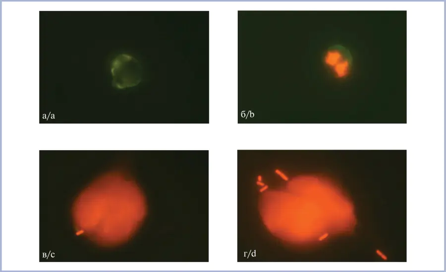
Направления исследований
ОБНАРУЖЕНИЯ НЕЙТРОФИЛЬНЫХ ВНЕКЛЕТОЧНЫХ ЛОВУШЕК В СУПРАВИТАЛЬНО ОКРАШЕННОМ ПРЕПАРАТЕ КРОВИ