
Лаборатория оптического биоимиджинга


Публикаций
87
Цитирований
3 045
Индекс Хирша
26
Необходимо авторизоваться.
Основным фокусом Лаборатории оптического имиджинга является создание технологий для оптического имиджинга, разработка новых подходов и алгоритмов автоматизации анализа имиджинговых данных, а также создание флуоресцентных и люминесцентных репортерных инструментов на основе генетических технологий. Имиджинг играет важную роль при визуализации физиологических и патологических процессов клеток в норме, а также их ответа на разрабатываемые прототипы лекарств, в том числе, противовирусные. Платформа включает в себя разработку генетически-кодируемых биомолекулярных сенсоров, используемых для изучения белок-белковых взаимодействий в живых клетках.
- Флуоресцентная микроскопия
- Конфокальная микроскопия
- Микроскопия сверхвысокого разрешения
- Получение бактериальных и эукариотических векторов с использованием генно-инженерных подходов
- Работа с культурами клеток растений, агротрансформация
- Клонирование Golden Gate
- Работа с культурами клеток млекопитающих, трансфекция
Всего публикаций
76
Всего цитирований
3014
Цитирований на публикацию
39.66
Среднее число публикаций в год
4.47
Годы публикаций
2008-2024 (17 лет)
h-index
26
i10-index
46
m-index
1.53
o-index
126
g-index
54
w-index
9
Описание метрик
h-index
Учёный имеет индекс h, если h из его N статей цитируются как минимум h раз каждая, в то время как оставшиеся (N - h) статей цитируются не более чем h раз каждая.
i10-index
Число статей автора, получивших не менее 10 ссылок каждая.
m-index
m-индекс ученого численно равен отношению его h-индекса к количеству лет, прошедших с момента первой публикации.
o-index
Среднее геометрическое h-индекса и числа цитирований наиболее цитируемой статьи ученого.
g-index
Для данного множества статей, отсортированного в порядке убывания количества цитирований, которые получили эти статьи, g-индекс это наибольшее число, такое что g самых цитируемых статей получили (суммарно) не менее g2 цитирований.
w-index
Если w статей ученого имеют не менее 10w цитирований каждая и другие статьи меньше, чем 10(w+1) цитирований, то w-индекс исследователя равен w.
Топ-100
Области наук
Журналы


Цитирующие журналы


Цитируемые журналы


Издатели
|
2
4
6
8
10
12
14
|
|
|
Springer Nature
13 публикаций, 17.11%
|
|
|
Pleiades Publishing
8 публикаций, 10.53%
|
|
|
Elsevier
7 публикаций, 9.21%
|
|
|
MDPI
7 публикаций, 9.21%
|
|
|
American Chemical Society (ACS)
5 публикаций, 6.58%
|
|
|
Royal Society of Chemistry (RSC)
5 публикаций, 6.58%
|
|
|
Public Library of Science (PLoS)
5 публикаций, 6.58%
|
|
|
openRxiv
5 публикаций, 6.58%
|
|
|
Wiley
4 публикации, 5.26%
|
|
|
International Union of Crystallography (IUCr)
2 публикации, 2.63%
|
|
|
Nizhny Novgorod State Medical Academy of the Ministry of Health of the Russian Federation
2 публикации, 2.63%
|
|
|
SPIE-Intl Soc Optical Eng
2 публикации, 2.63%
|
|
|
Taylor & Francis
1 публикация, 1.32%
|
|
|
Oxford University Press
1 публикация, 1.32%
|
|
|
Rockefeller University Press
1 публикация, 1.32%
|
|
|
EDP Sciences
1 публикация, 1.32%
|
|
|
Proceedings of the National Academy of Sciences (PNAS)
1 публикация, 1.32%
|
|
|
Frontiers Media S.A.
1 публикация, 1.32%
|
|
|
The Company of Biologists
1 публикация, 1.32%
|
|
|
eLife Sciences Publications
1 публикация, 1.32%
|
|
|
Anticancer Research USA Inc.
1 публикация, 1.32%
|
|
|
2
4
6
8
10
12
14
|
Организации из публикаций


Страны из публикаций
|
10
20
30
40
50
60
70
|
|
|
Россия
|
Россия, 62, 81.58%
Россия
62 публикации, 81.58%
|
|
США
|
США, 25, 32.89%
США
25 публикаций, 32.89%
|
|
Страна не определена
|
Страна не определена, 17, 22.37%
Страна не определена
17 публикаций, 22.37%
|
|
Великобритания
|
Великобритания, 8, 10.53%
Великобритания
8 публикаций, 10.53%
|
|
Германия
|
Германия, 6, 7.89%
Германия
6 публикаций, 7.89%
|
|
Австрия
|
Австрия, 6, 7.89%
Австрия
6 публикаций, 7.89%
|
|
Испания
|
Испания, 4, 5.26%
Испания
4 публикации, 5.26%
|
|
Япония
|
Япония, 3, 3.95%
Япония
3 публикации, 3.95%
|
|
Китай
|
Китай, 2, 2.63%
Китай
2 публикации, 2.63%
|
|
Швеция
|
Швеция, 2, 2.63%
Швеция
2 публикации, 2.63%
|
|
Франция
|
Франция, 1, 1.32%
Франция
1 публикация, 1.32%
|
|
Эстония
|
Эстония, 1, 1.32%
Эстония
1 публикация, 1.32%
|
|
Бразилия
|
Бразилия, 1, 1.32%
Бразилия
1 публикация, 1.32%
|
|
Израиль
|
Израиль, 1, 1.32%
Израиль
1 публикация, 1.32%
|
|
Финляндия
|
Финляндия, 1, 1.32%
Финляндия
1 публикация, 1.32%
|
|
Чехия
|
Чехия, 1, 1.32%
Чехия
1 публикация, 1.32%
|
|
Швейцария
|
Швейцария, 1, 1.32%
Швейцария
1 публикация, 1.32%
|
|
10
20
30
40
50
60
70
|
Цитирующие организации


Цитирующие страны
- Мы не учитываем публикации, у которых нет DOI.
- Статистика пересчитывается раз в сутки.
Направления исследований
Синтетическая биология агробактерий
+
Настоящий проект направлен на создание набора хорошо охарактеризованных генетических элементов для синтетической биологии агробактерий. Создание такого набора позволит быстро и модульно объединять различные репортерные и эффекторные генетические элементы в реплицирующиеся в агробактериях плазмиды с предсказуемой копийностью и уровнем экспрессии. Разработка набора с использованием современных подходов синтетической биологии позволит создать инструментарий, широко применимый для фундаментальных исследований этой важной группы бактерий. С его помощью можно будет также проводить точные исследования динамики и механизмов заражения растений агробактериями, механизмов интеграции геномной ДНК и взаимодействия с хроматином растений. Наконец, инструментарий для генетического редактирования агробактерий и изменения их физиологических процессов откроет возможности для создания новых штаммов с высокой эффективностью переноса ДНК в немодельные растения.
К/E-спирали: флуоресцентное мечение белков и наноскопия Protein-PAINT
+
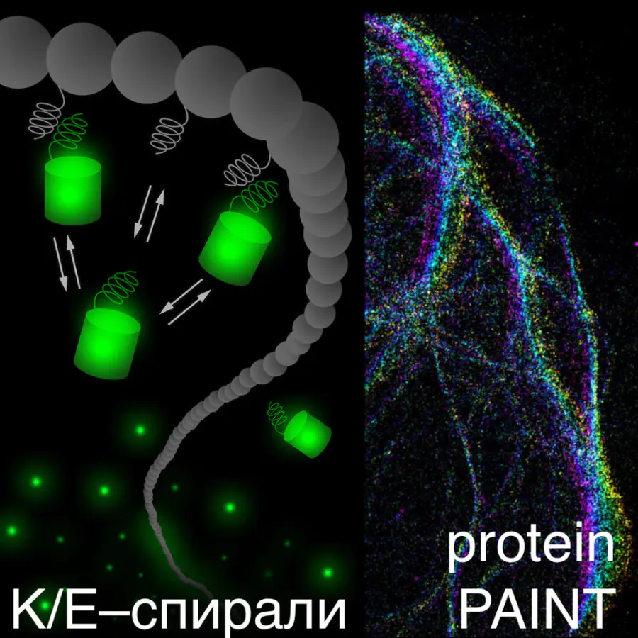
Одна из специально подобранных α-спиралей (K или E) служит меткой для целевого белка, а другую помещают на генетически кодируемый флуоресцентный белок. За счет обратимого взаимодействия K и E-спиралей целевая белковая структура окрашивается, а непрерывный обмен флуоресцентных белков в составе комплекса на порядок увеличивает устойчивость к фотообесцвечиванию. Небольшой размер меток (всего 2-3 кДа) позволяет сохранить нативную динамику исследуемых белков. Вдобавок, такой метод дает возможность наблюдать белки практически сразу после их синтеза. Наиболее интересным оказалось применение K/E-спиралей для Protein-PAINT – локализационной микроскопии сверхвысокого разрешения, основанной на обратимом взаимодействующих метках. Непрерывный обмен флуоресцентных белков между целевой клеточной структурой и цитоплазматическим пулом обеспечивает стабильно высокую плотность мечения даже при продолжительной съемке. С помощью K/E-спиралей впервые удалось воплотить концепцию наноскопии Protein-PAINT с использованием только генетически кодируемых репортеров.
Флуороген-активирующие белки, наноскопия и белок-белковые взаимодействия
+
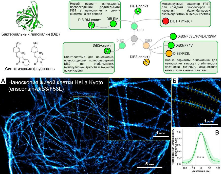
Создание меток на основе лиганд-связывающих белков и синтетических флуорогенов для использования в наноскопии живых клеток, а также в качестве модулируемых акцепторов FRET для изучения белок-белковых взаимодействий.
Публикации и патенты
Найдено
Ничего не найдено, попробуйте изменить настройки фильтра.

2022
—
2024
| Мишин Александр Сергеевич
2016
—
2018
| Мишин Александр Сергеевич
Партнёры


Адрес лаборатории
Москва, ул. Миклухо-Маклая, 16/10 к1
Необходимо авторизоваться.